Home

Flashy idiom Pronounce hyperintensity of spinal cord meaning statement precedent pea

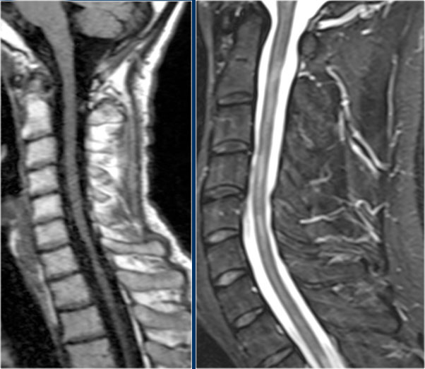
T1 Hyperintense Vertebral Column Melanoma Metastases | American Journal of  Neuroradiology
T1 Hyperintense Vertebral Column Melanoma Metastases | American Journal of Neuroradiology

Diagnostic Approach to Intrinsic Abnormality of Spinal Cord Signal  Intensity | RadioGraphics
Diagnostic Approach to Intrinsic Abnormality of Spinal Cord Signal Intensity | RadioGraphics

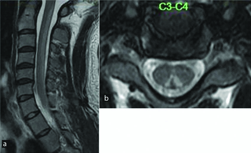
jcdr-13-TE01-g014.jpg

Diagnostic Approach to Intrinsic Abnormality of Spinal Cord Signal  Intensity | RadioGraphics
Diagnostic Approach to Intrinsic Abnormality of Spinal Cord Signal Intensity | RadioGraphics

Location, length, and enhancement: systematic approach to differentiating  intramedullary spinal cord lesions | Insights into Imaging | Full Text
Location, length, and enhancement: systematic approach to differentiating intramedullary spinal cord lesions | Insights into Imaging | Full Text

Differential diagnosis of T2 hyperintense spinal cord lesions: Part B -  Bou‐Haidar - 2009 - Journal of Medical Imaging and Radiation Oncology -  Wiley Online Library
Differential diagnosis of T2 hyperintense spinal cord lesions: Part B - Bou‐Haidar - 2009 - Journal of Medical Imaging and Radiation Oncology - Wiley Online Library

Diagnostic Approach to Intrinsic Abnormality of Spinal Cord Signal  Intensity | RadioGraphics
Diagnostic Approach to Intrinsic Abnormality of Spinal Cord Signal Intensity | RadioGraphics

Imaging of the spine and spinal cord: An overview of magnetic resonance  imaging (MRI) techniques - ScienceDirect
Imaging of the spine and spinal cord: An overview of magnetic resonance imaging (MRI) techniques - ScienceDirect

The Radiology Assistant : Myelopathy

Differential diagnosis of T2 hyperintense spinal cord lesions: Part B -  Bou‐Haidar - 2009 - Journal of Medical Imaging and Radiation Oncology -  Wiley Online Library
Differential diagnosis of T2 hyperintense spinal cord lesions: Part B - Bou‐Haidar - 2009 - Journal of Medical Imaging and Radiation Oncology - Wiley Online Library

Spinal cord T2-weighted MRI shows a hyperintense lesion extending from... |  Download Scientific Diagram
Spinal cord T2-weighted MRI shows a hyperintense lesion extending from... | Download Scientific Diagram

Intramedullary spinal tumor-like lesions - Edyta Maj, Katarzyna Wójtowicz,  Aleksandra, Podlecka-Piȩtowska, Marek Prokopienko, Andrzej Marchel, Olgierd  Rowiński, Monika Bekiesińska-Figatowska, 2019
Intramedullary spinal tumor-like lesions - Edyta Maj, Katarzyna Wójtowicz, Aleksandra, Podlecka-Piȩtowska, Marek Prokopienko, Andrzej Marchel, Olgierd Rowiński, Monika Bekiesińska-Figatowska, 2019

Advances in spinal cord imaging in multiple sclerosis - Marcello Moccia,  Serena Ruggieri, Antonio Ianniello, Ahmed Toosy, Carlo Pozzilli, Olga  Ciccarelli, 2019
Advances in spinal cord imaging in multiple sclerosis - Marcello Moccia, Serena Ruggieri, Antonio Ianniello, Ahmed Toosy, Carlo Pozzilli, Olga Ciccarelli, 2019

Spinal cord edema: unusual magnetic resonance imaging findings in cervical  spondylosis in: Journal of Neurosurgery: Spine Volume 99 Issue 1 (2003)  Journals
Spinal cord edema: unusual magnetic resonance imaging findings in cervical spondylosis in: Journal of Neurosurgery: Spine Volume 99 Issue 1 (2003) Journals
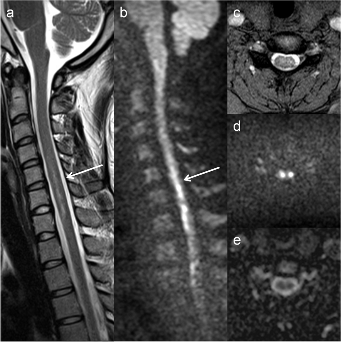
The utility of diffusion-weighted imaging in patients with spinal cord  infarction: difference from the findings of neuromyelitis optica spectrum  disorder | BMC Neurology | Full Text
The utility of diffusion-weighted imaging in patients with spinal cord infarction: difference from the findings of neuromyelitis optica spectrum disorder | BMC Neurology | Full Text
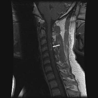
Spinal Cord - EMCrit Project

Multiple Sclerosis Spine Imaging: Practice Essentials, Computed Tomography,  Magnetic Resonance Imaging
Multiple Sclerosis Spine Imaging: Practice Essentials, Computed Tomography, Magnetic Resonance Imaging

Teaching NeuroImages: Spinal cord gray matter involvement in complex I  deficiency mitochondriopathy | Neurology
Teaching NeuroImages: Spinal cord gray matter involvement in complex I deficiency mitochondriopathy | Neurology

The Owl's Eyes Sign | Radsource

MRI spine showing hyperintensity in cervical cord C5 to C7 level (2) |  Download Scientific Diagram
MRI spine showing hyperintensity in cervical cord C5 to C7 level (2) | Download Scientific Diagram

Making Sense of MRIs for MS